
Immunohistochemistry
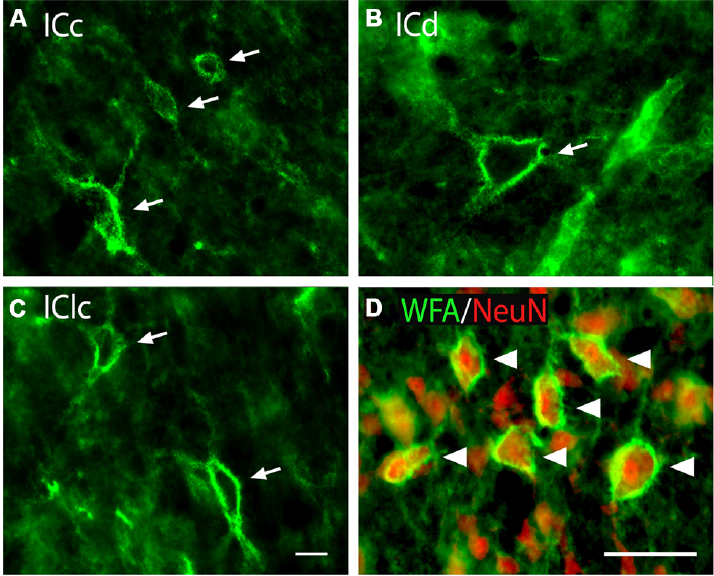
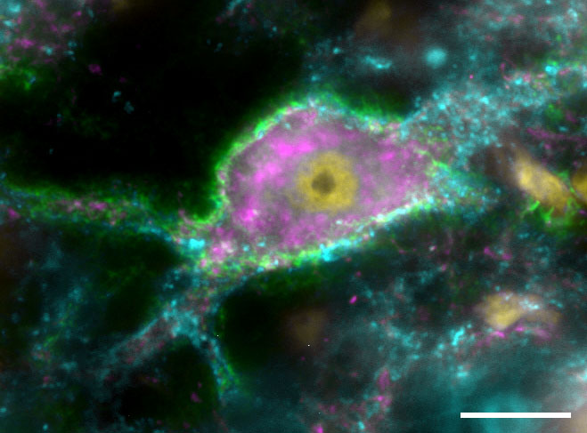
Immunostaining is a technique that uses antibodies to fluorescently label proteins. We also utilize lectin staining, Nissl staining, and others. Above, A lectin stain labels perineuronal nets (green), while an antibody to neuronal nuclear protein (NeuN, red) labels neurons.
Chemically-Selective Viral Tract Tracing
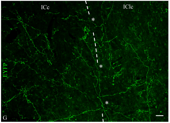
We combine transgenic animals with virally-induced fluorescent protein expression to trace selected populations of cells. Above, cre recombinase-dependent fluorescent protein genes were delivered to a midbrain cholinergic nucleus in rats expressing cre recombinase in cholinergic cells, allowing us to selectively label cholinergic axons from the PMT in auditory nuclei like the inferior colliculus.
Electron Microscopy
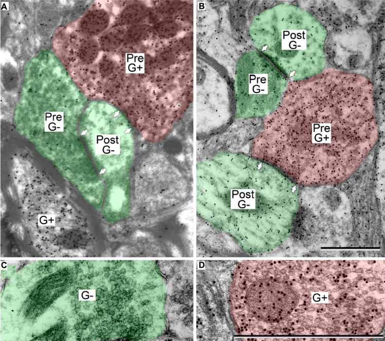
We use electron microscopy to examine structures at the sub-light level, including visualization of synapses. We can combine electron microscopy with post-embedding immunohistochemistry in order to examine the presence of various molecules. Above, GABAergic profiles (G+) were labeled with gold particles.
Fluorescent Tract Tracing
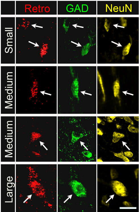
We use a variety of traditional anterograde and retrograde tract tracers to fluorescently or non-fluorescently label brain circuits associated with hearing. Above, retrograde tract tracers were deposited in the medial geniculate body and retrogradely-labeled cells in the inferior colliculus were examined.
Light Microscopy
We can use epifluorescence to image up to five fluorescent channels simultaneously. Each of our epifluorescent microscopes is equipped with an Apotome to allow for confocal-like images using structured illumination. We also use Neurolucida software to plot the distribution of cells of interest throughout the brain, and to reconstruct fluorescently-labeled neurons.
