
Our work has been funded through numerous sources, most prominently from the National Institute on Deafness and Other Communication Disorders. Our longstanding grant (NIH R01 DC004391), has been funded continuously since 1999. In 2020, we started a second grant (R01 DC018214) in collaboration with Dr. Michael Roberts at the University of Michigan. Below is a summary of the major questions we are addressing in these projects.
Modulatory Circuits in the Auditory System
R01 DC004391
What are the sources of excitatory or inhibitory cholinergic input to brainstem auditory nuclei?
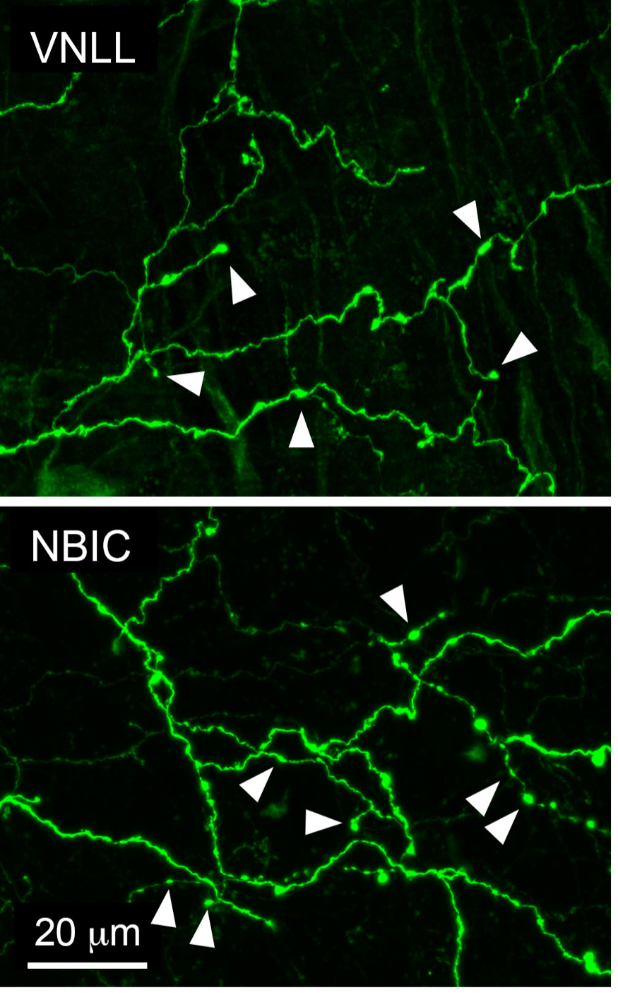
In the photos on the left, cholinergic axons are filled with a green fluorescent protein. Cholinergic axons with associated boutons can be seen in the ventral nucleus of the lateral lemniscus (VNLL) and the nucleus of the brachium of the inferior colliculus (NBIC), two areas where cholinergic inputs are poorly understood. Arrowheads indicate boutons, which are likely sites of neurotransmitter release. We are currently using viral tract tracing in a newly developed mouse model to determine the sources of this cholinergic input, and we will use electron microscopy to determine whether cholinergic synapses in these and other brainstem nuclei are excitatory or inhibitory.
Do cholinergic cells simultaneously innervate multiple auditory nuclei?
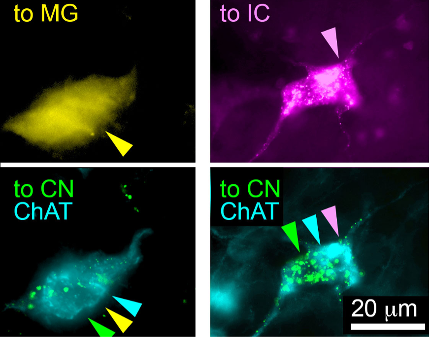
The left panel of the photo on the left shows a neuron that projects simultaneously to the medial geniculate body (MG, yellow), and the cochlear nucleus (CN, green). The right panel shows a neuron that simultaneously projects to the inferior colliculus (IC, magenta) and the cochlear nucleus (green). Both neurons use acetylcholine as a neurotransmitter (cyan). We are currently using fluorescent tract tracing to ask how often cholinergic cells have branching axons to multiple auditory targets, which provides an opportunity for simultaneous release of acetylcholine across auditory nuclei.

What types of cholinergic receptors are associated with ascending or descending auditory circuits?
In the photo on the left, cells in the dorsal part of the inferior colliculus (ICd) have been retrogradely filled via injections into the medial geniculate body (MG, red). These cells also express mRNA for the cholinergic M2 receptor, a type of muscarinic receptor (cyan). We will use a technique called in-situ hybridization to look at the expression of various types of cholinergic receptors within fluorescently-labeled auditory pathways.
Which cholinergic cells are targeted by projections from the auditory cortex, and where do those cholinergic cells project?
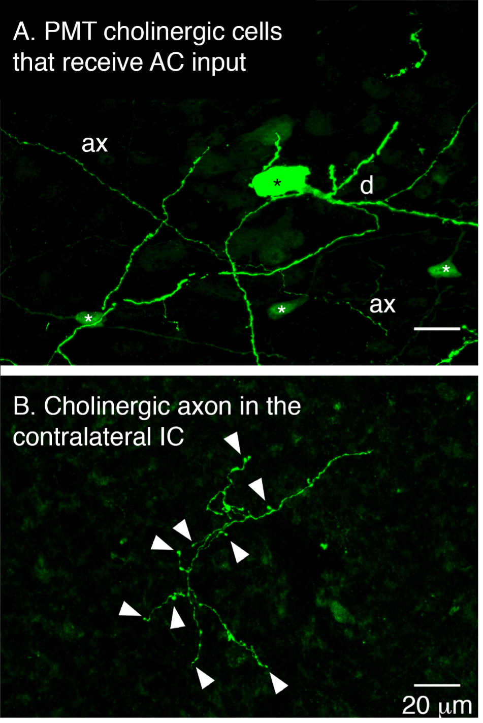
In the top photo on the left, a cholinergic cell in the pontomesencephalic tegmentum (PMT) has been trans-synaptically labeled with a green fluorescent protein via a viral injection in the auditory cortex (AC), indicating that this cell receives synaptic input from the auditory cortex. Both dendrites (d) and axons (ax) of the cell are visible, including axons which project to other auditory nuclei. On the bottom, an axon from a similarly-labeled PMT cell that receives auditory cortex input can be seen in the inferior colliculus (IC), indicating release of acetylcholine in the inferior colliculus that may be driven by the auditory cortex. We will use this technique in addition to more traditional tract-tracing techniques to identify nuclei that get cholinergic input from cells targeted by the auditory cortex.
Circuit Mechanisms for Auditory Processing in the Inferior Colliculus
R01 DC018214
What are the inputs and outputs of two subclasses of cells in the inferior colliculus?
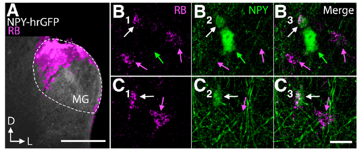
In the photo above, inferior colliculus cells that express neuropeptide Y (NPY, green) are retrogradely labeled after an injection of retrograde tracer into the medial geniculate nucleus (magenta), meaning that NPY cells can send projections to the medial geniculate nucleus. We have also shown projections from NPY cells within the ipsilateral inferior colliculus and to the contralateral inferior colliculus. We are currently working to address the possibility that NPY cell may also project to the nucleus of the brachium of the inferior colliculus. This project also addresses the circuitry of inferior colliculus cells that express vasoactive intestinal peptide, which we’ve shown have widespread projections.
Past and Present Funding
National Institutes of Health
1999–present R01 DC004391 Modulatory Circuits in the Auditory System
2020–present R01 DC018214 Circuit Mechanisms for Auditory Processing in the Inferior Colliculus
2019–present R01 DC016918 Neuronal Hyperactivity: Tinnitus and Distress (PI: A. Galazyuk)
2016–2019 R01 EY026662 Metabolic Vulnerability as a Disease Target for Glaucoma (PI: D. Inman)
2012–2017 R01 EY 022358 Axonopathy in Glaucoma (PI: S. Crish)
1998–1999 R55 DC003790 Functional Anatomy of the Auditory Pathways
NIH National Research Service Awards to lab trainees
2014-2016 F31 DC014228 Predoctoral NRSA (PI: Nichole Foster [Beebe])
2012–2015 F32 DC012450. Postdoctoral NRSA (PI: Jeffrey Mellott)
2010–2012 F32 DC010958. Postdoctoral NRSA (PI: Kyle Nakamoto)
2007–2009 F31 DC08463. Predoctoral NRSA (PI: Susan Motts)
2001–2004 F31 DC05277. Predoctoral NRSA (PI: Diana Coomes Peterson)
NEOMED Research Incentive Program
2006–2007 Physiology and Morphology of Guinea Pig Auditory Cortex Neurons (Co-investigator: Y. Lu)
2009–2010 Modulation of Synaptic Transmission in the Prefrontal Cortex by the Tachykinin NK3 Receptor (Co-investigator: M. Simmons)
Deafness Research Foundation 1997–1998 Research Grant
